Mole Removal
What is a mole?
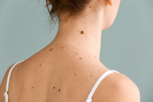
Moles, medically known as mole-nevi are growths on the skin that are generally brown or black and situated either alone or in groups on the body. Most people have them, with the average person having 10- 40 moles by adulthood. They are generally no cause for concern unless they change size, colour or shape.
Moles can develop anywhere on the body and vary in shape, colour and texture. Some are small, smooth and round or oval with others appearing raised or with hairs in them.
As the years pass, it is normal for moles to change slowly, for new moles to appear or for them to fade and disappear entirely. During pregnancy or in sunlight exposure, you may notice moles get slightly darker.
Different types of moles
Congenital Nevi
Congenital nevi are moles that exist at birth and appear in about 1 in 100 people. These moles are more likely to develop into melanoma (cancer) than moles that occur after birth.
Dysplastic Nevi
Dysplastic nevi are moles that are generally larger than average and abnormal in shape with the colour tending to be uneven with dark brown centres and lighter edges. These moles are generally hereditary with people who have 10 or more on their body, having a higher chance of developing melanoma (cancer). There are numerous types of moles, organised by their appearance when they develop, and their risk of becoming cancerous. It is important that any changes in a mole are checked by a Dermatologist.
What causes a mole?
Moles develop when cells in the skin grow in a group, instead of being spread throughout the skin. These cells are called melanocytes, and they make the pigment that gives skin its natural colour.
Moles typically develop in childhood and adolescence and change in size and colour as you mature. New moles usually appear at times when your hormone levels change, such as during pregnancy.
A new mole may appear due to a number of reasons, including:
- Aging
- Family history of developing unusual moles
- Having fair skin and fair or red hair
- Response to medications such as antibiotics, hormones, antidepressants or immune system suppressing drugs
- Genetic mutations
- Sunburn, sun exposure, or sunbed use
Symptoms
Moles can be a valuable sign to alert you of the onset of cancer. If they change shape and colour this can be a sign of a melanoma beginning to form. Melanoma is the most prevalent form of cancer in young women. The most common area for melanoma to be found in men is the chest, and back and in women, it is the lower leg.
The ABCDE guide can help you determine important characteristics when examining moles. If a mole shows any of the signs listed below, get it checked immediately by a Dermatologist.
Asymmetrical shape. One half is dissimilar to the other half.
Border. Look for moles with uneven, patchy borders.
Colour. Look for growths that have changed colour, have various colours or have uneven colour.
Diameter. Look for new growth in a mole greater than 1/4 inch (about 6 millimetres).
Evolving. Watch for moles that change in colour, size, shape or height. Moles may also develop new symptoms, such as itchiness or bleeding.
If you notice any of the above, please book an appointment to see your GP regarding mole removal.
Prevention
Watching for changes, looking after and paying attention to your skin are all steps you can do to reduce the development of moles and the risk of melanoma.
Regularly examining your skin by using a mirror to see the parts of your body you cannot easily reach will help you become familiar with the patterns, locations and characteristics of your moles. Try protecting your skin from UV radiation by avoiding peak sun times, sunbeds and wearing a minimum of SPF 30 all year round. The sunscreen should preferably have a 5* rating.
Diagnosis
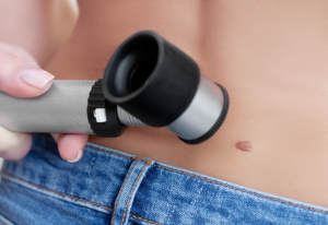
Your Dermatologist can identify moles by examining your skin. If they suspect that the mole may be cancerous or cause for concern, they will carry out a skin biopsy, (performed under local anaesthetic numbing the affected area), where a small sample of the mole is taken to examine under a microscope. A diagnosis can typically be made in less than a week.
A Dermatologist uses a skin biopsy to remove cells or skin samples from the surface of your body to be examined, diagnosed and to provide evidence about your medical condition.
The main type of skin biopsies:
Shave Biopsy
A doctor uses a sharp blade similar to a razor to remove a thin section of skin punch biopsy. A doctor uses a circular tool to remove a tube-shaped section of skin, including deeper layers.
Excisional Biopsy
A doctor uses a scalpel to remove a whole lesion or area of irregular skin, including a section of normal skin down to the fatty layer.
Other tests you may have include a CT scan or MRI scan.
If the mole is found to be cancerous, it will require a second operation to be completely removed, ensuring no cancerous cells are left in the skin. Further tests may also be required if it is feared that cancer has spread into your blood, bones or other organs. Even if your mole is not cancerous, you may still decide to have it removed for cosmetic reasons.
Treatment and procedure
We offer mole removal for both cosmetic and medical reasons. Moles are most commonly removed for medical reasons if your Dermatologist suspects they might contain abnormal or cancerous cells. Most mole removals are usually carried out on an outpatient basis. Your doctor numbs the area around the mole using local anaesthetic and removes it, along with a section of healthy skin if essential; this may leave a permanent scar.
Your GP will refer you to a Dermatologist if any of your moles are suspected of containing abnormal or cancerous cells; these will be removed and sent to be checked for cell content. in a procedure called a biopsy.
The procedure chosen to remove your mole will depend on the type of mole you have. The dermatologist will advise which technique is most suitable for you.
Recovery
Recovery from the mole removal procedure is relatively quick. You will be able to carry out your usual activities the following day (unless your work puts strain on your stitches). The scar should heal within 2-3 weeks, and any pain should be mild and treatable with over-the-counter relief. Occasionally your doctor will ask you to return after your procedure to check the wound and change the dressing; you may also need to have your stitches removed if they are not dissolvable.
Complications
It is unusual for this procedure to restrict any daily activities; however, all surgical procedures include a certain degree of risk and can result in complications. These include:
- Pain that is usually mild and can be treated with over-the-counter pain killers
- Bleeding (blood clots or excessive bleeding)
- Infection of the wound that results in redness and swelling. This can be treated with antibiotics
- Scarring, although this usually fades over time, although there is likely to be a permanent mark on your skin
- Wound healing problems (there is a higher risk if you have other skin conditions)
- The mole may come back. A common mole will not return after it has been removed, but a cancerous one might
If you develop any problems following your mole removal, you should speak to your Dermatologist so you can get any additional care that you need.
Outlook Following Mole Removal Surgery
Mole removal surgery is typically successful and you will not require any further treatment. However, there is a chance that you will need additional surgery if the mole starts to grow back. You might also need extra tests or treatment if cancer cells are found when the mole is tested. For example, a larger area of skin may need to be removed and tested if there is a possibility that some cancerous skin cells have been left behind.
You can use your private medical insurance or pay for your Mole Removal treatment. We offer competitive, fixed price packages. If you are using your health insurance, please contact your insurer first for approval and let them know you’d like to be treated at One Hatfield Hospital.
Why One Hatfield
- Modern purpose-built hospital opened in December 2017
- 0% and low finance options**
- Fast access to diagnostics including MRI, X-ray and Ultrasound
- Private, spacious, en-suite rooms
- Specialist physiotherapy and nursing teams
- Little or no waiting time
- ‘Ultra clean air’ theatres
- Freshly prepared food
- Calm, dignified experience
**Terms and conditions apply
Contact us and find out more
If you are based in and around Hertfordshire, St Albans, Stevenage, Watford, Barnet, North London, Welwyn or Bedfordshire and would like to visit the One Hatfield Hospital please click here.
Dermatology Pricing Guide at One Hatfield Hospital
This is a list of guide prices for some of common Dermatology treatments and procedures.
| Treatment | Guide Price |
|
Incision of lesion (1 -3) |
£660* |
|
Incision of lesion (4 -7) |
£945* |
|
Cryotherapy (wart removal) |
£100* |
| PRP Hair loss injections | £370.30 |


 One Ashford
 One Hatfield